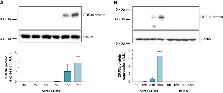
Figure 1.
Time course of ORF 3a protein expression in SARS-CoV-2-infected human-induced pluripotent stem cell-derived cardiomyocytes (hiPSC-CMs) and human cardiac fibroblasts (hCFs). (A) Human-induced pluripotent stem cell-derived cardiomyocytes were subjected to SARS-CoV-2 infection [multiplicity of infection (MOI) of 1.0], and cells were harvested at various time points post-infection as specified. The upper section shows representative immunoblot signals for ORF 3a protein and β-actin as loading control. The lower section depicts the average ORF 3a protein signals, obtained from n = 3 independent experiments, normalized to β-actin and presented in arbitrary units (A.U.). (B) Both hiPSC-CMs and hCFs were exposed to SARS-CoV-2, and cells were harvested at designated time intervals. The top section illustrates the immunoblot signals for ORF 3a protein and β-actin from the respective cell lysates. The bottom section presents the average ORF 3a protein signals from n = 3 experiments, adjusted relative to β-actin expression and measured in A.U. Statistical significance is indicated by ***, representing a P < 0.001 from a two-tailed unpaired Student’s t-test. Data are expressed as mean ± standard error of the mean.

