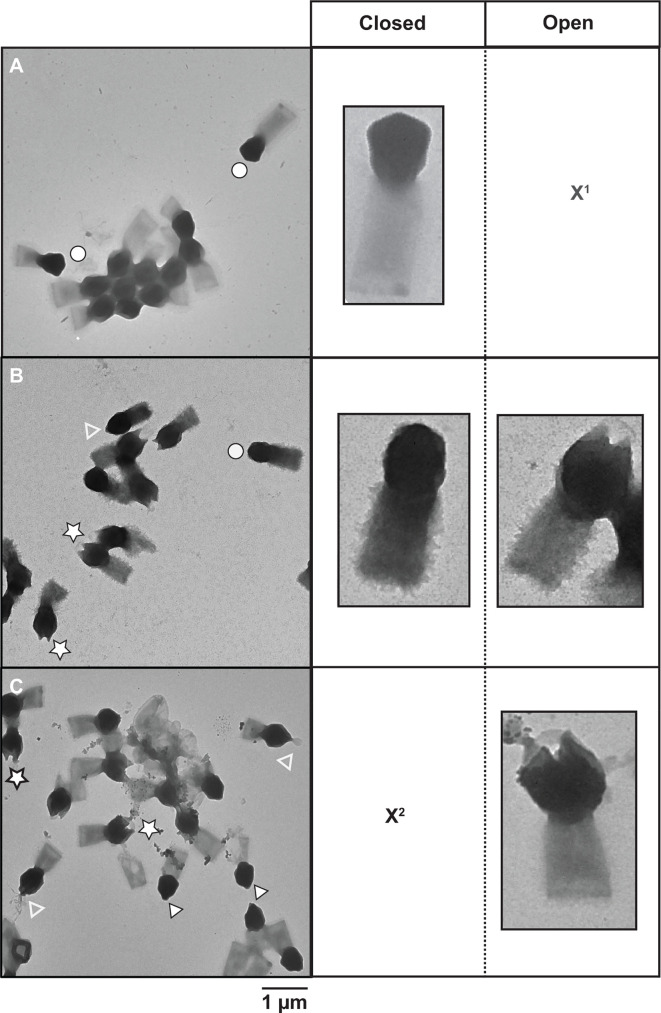
Fig 2.
Effect of copper and iron ions on TPV capsids. TEM images of (A) untreated TPV, (B) 10 mM CuCl2-treated particles, and (C) 10 mM FeCl3-treated particles. Images depict the three stages of stargate features: closed (white circles), pre-opened (white arrowheads), and open (white stars). The empty white arrowheads point at what we hypothesize to be the viral seed exiting the interior of the capsids. X1: open particles not present in this view; X2: closed partciles not present in this view. Scale bar: 1 µm.

