Fig 3.
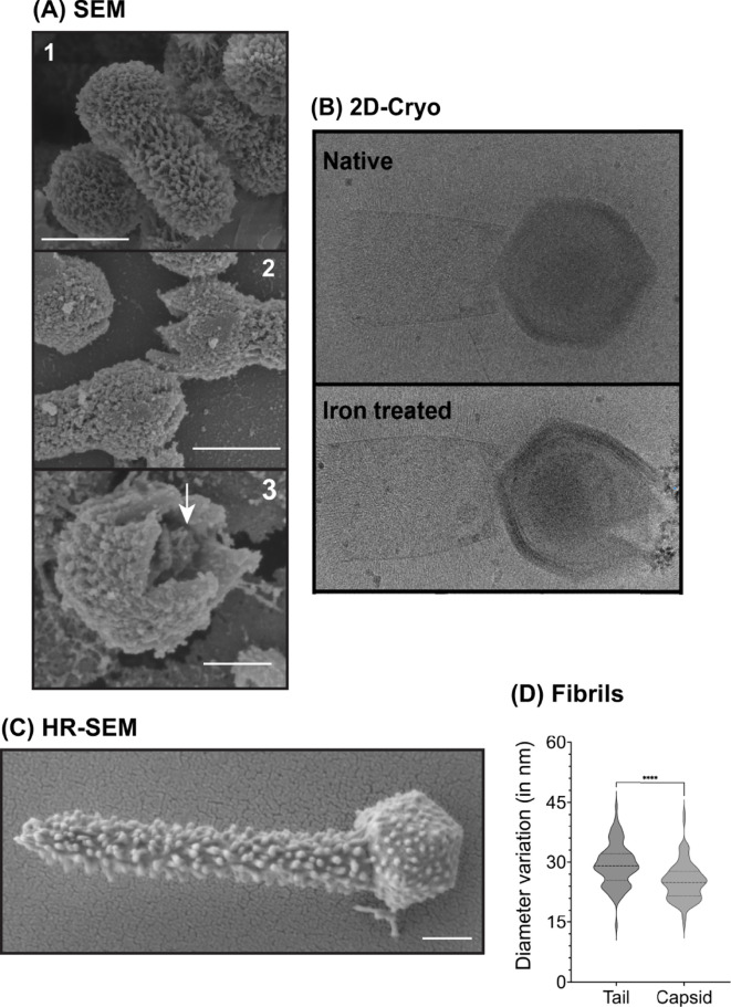
Structural features of TPV particles under native and metal-treated conditions. (A) Scanning electron microscopy images of native or copper-treated TPV. Images show (1) the closed, native virus particles; (2) intermediate steps on TPV stargate opening; and (3) the interior of the capsid, where we hypothesize the genome sac is still present even after copper treatment (indicated by the white arrow). Scale bar: 0.5 µm. SEM was not amenable to iron-treated particles, as this metal accumulates on the particle surface. (B) 2D cryo-EM images of TPV under native (closed particle, top panel) or iron treated (open stargate, bottom panel); white arrows indicate the genome sac; scale bar: 0.5 µm. (C) Ultrahigh-resolution TEM of a native (untreated) TPV particle. In this image, the fibrils are the main feature on the particle surface. The virion shows the tail at its upper-end length, ~1.4 µm. Scale bar: 0.2 µm. (D) Based on C, fibrils were measured: on average, capsid fibrils measured 25.14 nm, and tail fibrils, 29.29 nm.

