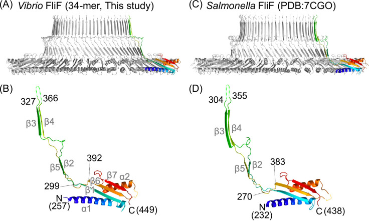
Fig 2.
Comparison of the S-ring and FliF structures of Vibrio and Salmonella. (A) Cross-section view of the Vibrio S-ring structure shown as a Cα ribbon drawing. A protomer is colored in rainbow gradient from blue at the N-terminus to red at the C-terminus. (B) Magnified view of the protomer in panel A. (C) Cross-section view of the Salmonella S-ring structure. (D) Magnified view of the protomer in panel C. The secondary structure elements are labeled with gray text in panels B and D.

