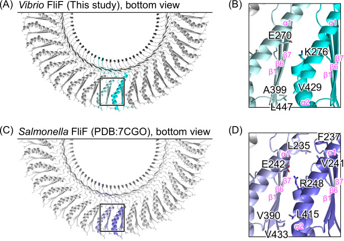
Fig 4.
Comparison of the inter-subunit interface in the S-ring of Vibrio and Salmonella. (A and C) Bottom view of the Cα ribbon drawing of the S-ring structures of Vibrio and Salmonella. (B and D) Magnified view of the squared region in panels A and C. The side chains contributing to the inter-subunit interaction in the RBM3 are shown in stick models. The secondary structure elements are labeled with pink text in panels B and D.

