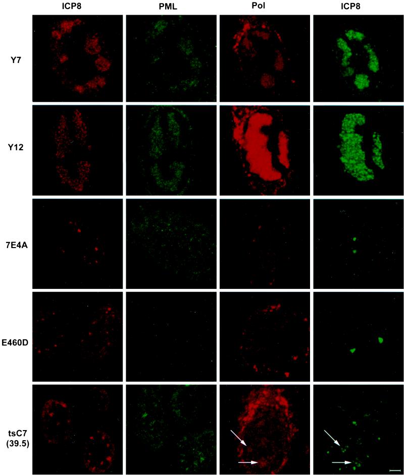
FIG. 5.
Localization of ICP8, PML, and Pol in cells infected with various Pol mutant viruses. Cells were infected with wild-type or mutant HSV-1 for 6 h and double stained with antibodies against the major viral DNA-binding protein ICP8 and either the cellular ND10 protein PML (PG-M3) or the viral polymerase protein (M1). The first two columns depict the same cell labeled with antibodies recognizing ICP8 (red) or PML (green); the second two columns depict a different cell colabeled with antibodies to ICP8 (green) and Pol (red). Y7 and YD12 represent the mutants that are viable on Vero cells. 7E4A represents the mutants that made no Pol that was detectable by Western blotting. E406D represents the mutants that made Pol that was detectable by Western blotting but did not show Pol localization to prereplicative sites. At the nonpermissive temperature of 39.5°C, tsC7 made Pol that was detectable by Western blotting and showed weak localization of Pol to accumulations of ICP8 (arrows); this phenotype is seen in PAA-treated KOS-infected cells (not shown). The cytoplasmic staining observed in these experiments may be due to secondary antibody binding of the viral Fc receptors in the endoplasmic reticulum and Golgi apparatus of infected cells. Bar, 10 μm.

