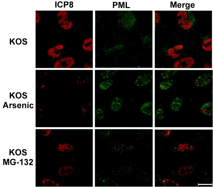
FIG. 8.
Localization of ICP8 and PML in cells treated with arsenic or MG-132. HEp-2 cells were infected with KOS at an MOI of 10 for 6 h in the presence or absence of 10−6 M As2O3 or 0.05 mM MG-132. Cells were pretreated with drug for 30 min prior to infection. After fixation, cells were stained for ICP8 (shown in red) and PML (shown in green). Bar, 25 μm.

