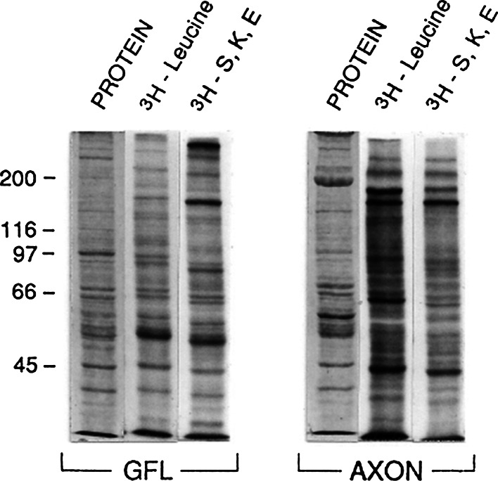
Fig. 3.
SDS-PAGE separations of radioactive proteins incorporated into and extracted from GFL and ensheathed giant axons after incubation of tissues in either 3H-leucine or 3H-serine, 3H-lysine, and 3H-glutamate (3H–S, K, E) mixtures. The lanes labeled PROTEIN illustrate the Commassie blue staining patterns of proteins in the GFL and giant axon extracts, and the numbers to the left represent the migration positions the proteins of known molecular weights (kD × 10E−3). The radioactive proteins in these extracts were also used in the immunoprecipitation experiments illustrated in Figs. 4 and 5

