Figure 2.
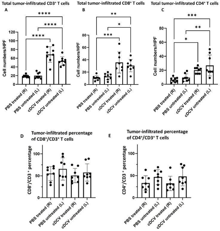
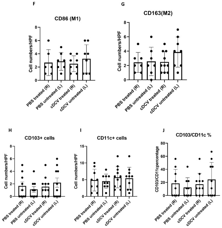
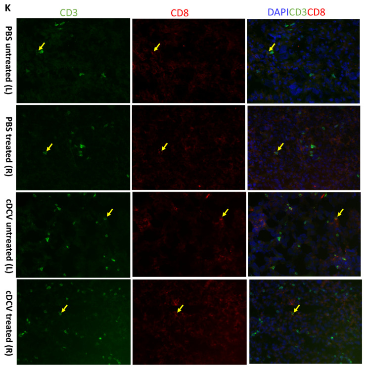
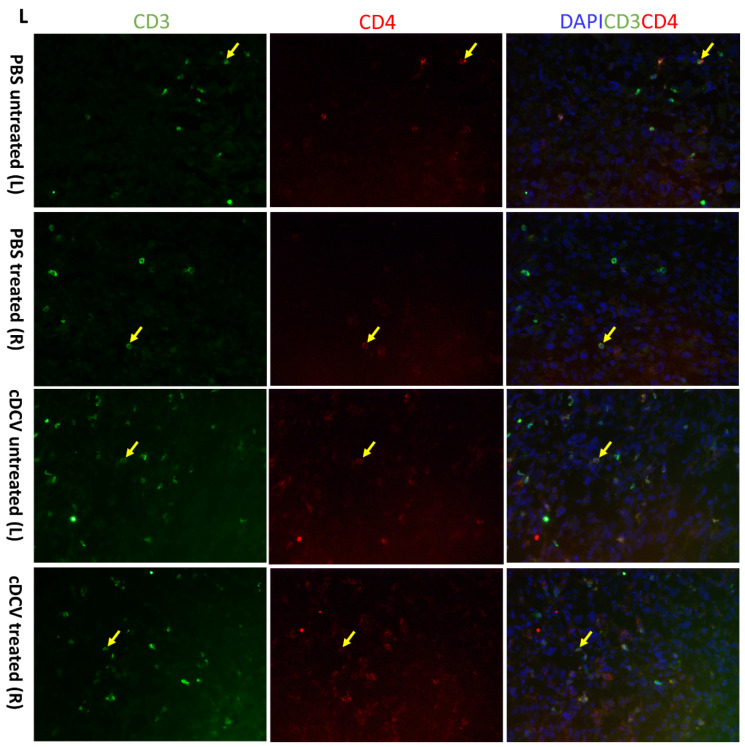
Effect of cDCV treatment on infiltrating T-cells and macrophages. Bilateral tumors were stained with anti-CD3/anti-CD8/anti-CD4 (A–C,K,L), anti-CD11c/anti-CD103 (H,I), anti-CD86 (F), or anti-CD163 (G) antibodies. Percentage of CD8+ or CD4+ cells out of CD3+ cells were showed in (D,E). Percentage of CD103+ cells out of CD11c+ cells was showed in (J). Positive cells were quantified using Simple PCI software. n = 7–8 mice per group. Data were analyzed using one-way ANOVA test. * p < 0.05, ** p < 0.01, *** p < 0.001, **** p < 0.0001. cDCV: type 1 CD103+ dendritic cell vaccine; HPF: high-power field; L: left side; PBS: phosphate-buffered saline; R: right side. All values reported represent means ± SEMs. Arrows indicates an example of a positive stained cell in each field.


