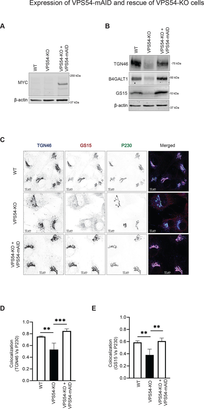
Figure 1: Expression of VPS54-mAID rescues VPS54-KO defects.

(A) Western blot (WB) analysis of RPE1 cell lysates from wild-type (WT), VPS54 knock out (VPS54-KO), and VPS54-KO cells rescued with VPS54-mAID. Blots were probed with anti-myc (to detect VPS54-myc-mAID) and anti-β-actin antibodies. (B) WB analysis of RPE1 cell lysates from WT, VPS54-KO, and VPS54-mAID, probed with anti-TGN46, anti-B4GALT1, and anti-GS15 antibodies. β-actin was used as a loading control. (C) Confocal microscopy images of WT, VPS54-KO, and VPS54-mAID RPE1 probed for TGN46, GS15, and P230. (D) Quantification of IF images in (C). Pearson’s correlation coefficient was used to assess the colocalization of TGN46 and P230. (E) Pearson’s correlation coefficient was used to assess the colocalization of GS15 and P230. At least 50 cells were analyzed per sample for the quantification. Statistical significance was determined using one-way ANOVA. ** p≤ 0.01, *** p ≤ 0.001.
