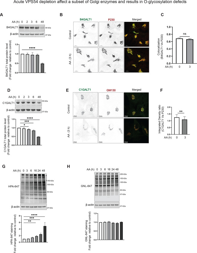
Figure 5: Acute VPS54 depletion affects a subset of Golgi enzymes and results in O-glycosylation defects.
(A) WB analysis of cell lysates of AA treated RPE1 VPS54-mAID cells probed with (top panels) anti-B4GALT1 (A), and anti-C1GALT1 (D). β-actin was used as a loading control. The bottom panels on (A), and (D) are the quantification of the blots from three independent experiments. (B) Airyscan microscopy of RPE1 VPS54-mAID cells untreated (control) or treated with AA for 3 h and co-stained for B4GALT1 and P230. (C) Colocalization of B4GALT1 with P230 was determined by calculation of the Pearson’s correlation coefficient. (E) Airyscan microscopy of RPE1 VPS54-mAID cells untreated (control) or treated with AA for 3 h and co-stained for C1GALT1 and P230. (F) Integrated density ratio of C1GALT1 to P230 in control and AA treated group was determined using ImageJ. Statistical significance was calculated using paired t-test. ** p≤ 0.01. (G)Total proteins from AA treated RPE1 VPS54-mAID were resolved by SDS-PAGE and probed with (Top panel) HPA-647 (G), and GNL -647 (H). The bottom panels on (G), and (H) are the quantification of the blots from three independent experiments. Statistical significance was calculated using one-way ANOVA. ** p≤ 0.01, *** p≤ 0.001, **** p≤ 0.0001.

