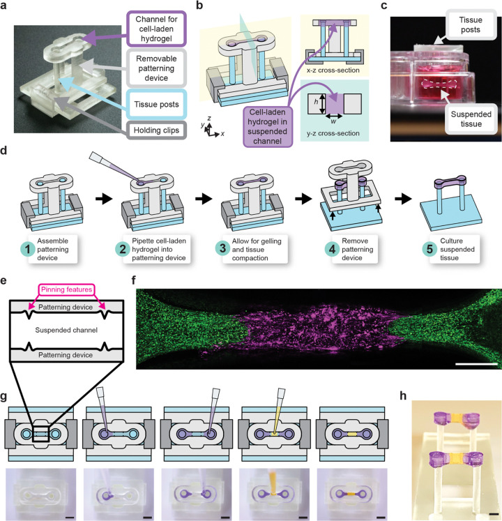
Figure 1. Workflow of generating single and multi-region suspended tissues using the STOMP platform.
a) Image of the STOMP platform, which includes a removable patterning device containing an open channel that interfaces with a pair of vertical posts. The patterning device is held in place to the base for the posts with holding clips. b) Schematic of the STOMP platform. A cell-laden hydrogel is pipetted into the open channel, where it flows via surface-tension driven forces across the open channel and anchors onto the suspended posts, thus generating a free-standing suspended tissue. c) Side view of the resulting suspended tissue cultured in a 24-well plate. d) Workflow of patterning a tissue composed of a single region, where the composition is the same across the tissue. e) Top-down view of the capillary pinning features along the open channel that are used to pin the fluid front. f) Fluorescent image of patterned 3T3 mouse fibroblast cells laden in a fibrin hydrogel using STOMP. The outer region of 3T3 cells were dyed by CellTracker Green (green) and were pipetted first. The middle region of 3T3 cells were dyed by CellTracker Red (magenta). Scale bar is 500 μm. g) Workflow of patterning tissues comprising three distinct regions. Corresponding video stills show patterning of purple-colored agarose in the outer regions first, followed by patterning yellow-colored agarose in the middle region. Full video can be seen in Supplementary Video 1. All scale bars are 2 mm. h) Side view image of multi-region agarose suspended hydrogel construct. Scale bar is 2 mm

