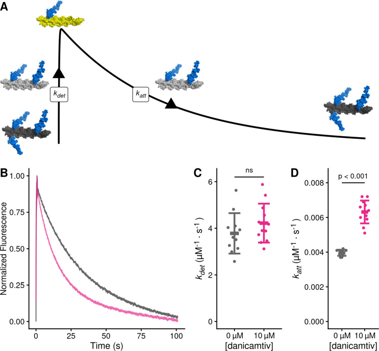
Figure 5. Danicamtiv increases myosin’s attachment rate in a single turnover stopped flow assay.
Black = DMSO control. Pink = 10 μM danicamtiv. A) Schematic conceptually describing the single turnover assay used to measure myosin’s attachment and detachment rates. In this assay, myosin and pyrene labeled actin are pre-incubated and then mixed with a sub-saturating concentration of ATP. The pyrene fluorescence increases as myosins detach from actin, and the increase in fluorescence reports the rate of detachment of myosin from actin (kdet). Myosin then reattaches to actin, quenching the fluorescence and reporting the attachment rate (katt). B) Fluorescence transients from the single turnover assay. Data were fitted as described in the Supplemental Methods. C) The average second-order rate of detachment (kdet) was similar with and without 10 μM danicamtiv (3.8 ± 0.9 vs. 4.2 ± 0.8 s−1; P = 0.23). D) The second-order rate of attachment (katt) increased with the addition of 10 μM danicamtiv (0.0040 ± 0.0002 vs 0.0063 ± 0.0007 μM−1·s−1; P = < 0.001). For C and D, the thick lines show the average values, and the error bars show the standard deviation. The individual points are the fitted second-order rates to individual transients collected across three experimental replicates. Statistical testing was done using a two-tailed T-test after passing a normality test.

