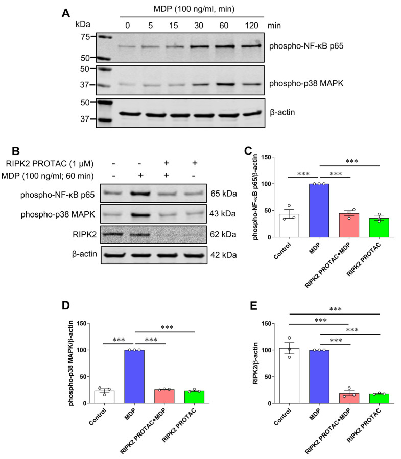
Figure 4. RIPK2 PROTAC suppresses activation of both NF-κB p65 and MAPK p38 induced by MDP in SIM-A9 cells.
A) SIM-A9 cells were stimulated with 100 ng/mL MDP for the indicated periods (0 to 120 min). Immunoblots show MDP increased phosphorylation of NF-κB p65 and MAPK p38, and 60 min incubation of MDP induced the maximal effects on both phosphorylated protein levels. Data are representative of three independent experiments with similar results. B-E) SIM-A9 cells were pretreated with 1 μM RIPK2 PROTAC for 4 h followed by 60 min incubation of 100 ng/mL MDP. After that, cells were harvested for protein extraction and western blots. Immunoblots and graphs show that RIPK2 PROTAC completely abolished the effects of MDP on the phosphorylation of both NF-κB p65 (B, C) and MAPK p38 (B, D), which was associated with marked degradation of RIPK2 by its PROTAC pretreatment (B, E). One-way ANOVA with Bonferroni post-test, ***P<0.001. Data are normalized to β-actin and represented as mean ± SEM from three independent experiments.

