Figure 1:
Assembly and structural analysis of liposome-templated HIV-2 CLPs
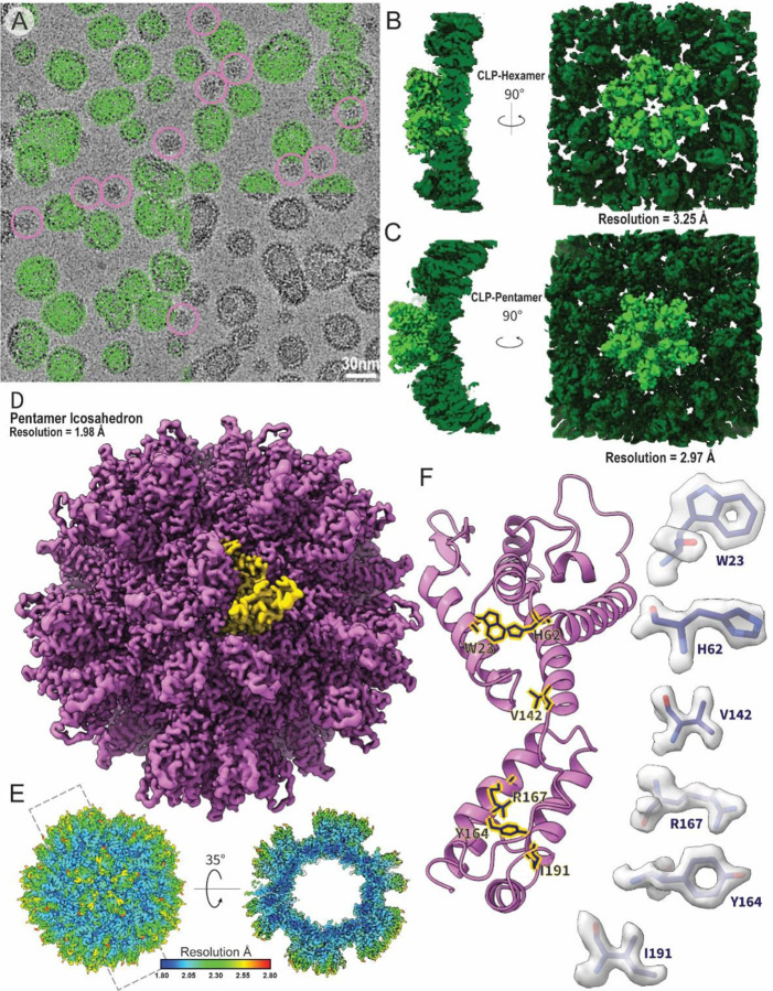
A. Example cryo-EM micrograph of liposome-templated HIV-2 CLPs. Green circles mark example particles picked for 3D reconstruction. Pink circles mark example particles picked for icosahedral assemblies. B. Cryo-EM map of the HIV-2 CA hexamer. C. Cryo-EM map of the HIV-2 CA pentamer. D. Cryo-EM map of the micelle-templated HIV-2 pentamer icosahedron. Yellow highlights a single CA monomer. E. Local resolution map of the micelle-templated HIV-2 pentamer icosahedron. F. Cartoon representation of a CA monomer from the pentamer icosahedron. Sticks correspond to selected residues with corresponding map density highlighted to the sides.

