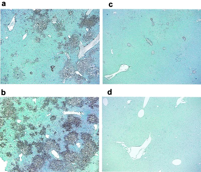
FIG. 4.
Immunohistochemistry of liver sections of C57BL/6 mice infected with the recombinant viruses SA59R13 (a), Penn98-1 (b), and S4R21 (c) and a mock-infected mouse (d). Mice were infected as described for Fig. 2 and then sacrificed at day 5 p.i. Livers were removed, fixed and sectioned. MHV was detected by immunolabeling with an MAb against the nucleocapsid (N) protein of MHV, using the avidin-biotin-immunoperoxidase technique (Vector) as described in the text. Viral antigen was associated with areas of hepatocellular necrosis in SA59R13 (a) and Penn98-1 (b). S4R21-infected mice (c) showed low levels of viral antigen staining. No signs of pathology or viral antigen were found in a mock-infected control (d). Magnification, ×40.

