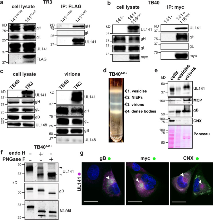
Fig. 1. UL141 is incorporated into virions and assembles into a gH/UL116 complex.
a, The HCMV BAC-derived strain TR3 was modified to introduce a FLAG tag at the C-terminus of UL141 (TR3–141F). Infected fibroblasts were lysed 72 h post infection (hpi), and IP was performed with anti-FLAG. IP eluates were resolved by SDS-PAGE and analyzed by immunoblot with the indicated antibodies. b, Fibroblasts were infected with BAC-derived TB40/E (TB40-BAC4) repaired for UL141 expression and engineered to express a myc-tagged UL116 (141+,116myc). Anti-myc IP was carried out at 72 hpi and eluates were analyzed by Western blot. c, Infected cell lysates and purified virions from HCMV strains TB40 (141-) and TR3 (141+) were compared for the indicated viral glycoproteins. d, TB40–141+ viruses were grown in fibroblasts and concentrated through a 20% sorbitol cushion prior to glycerol-sodium tartrate gradient purification to separate infectious virions (band 3) from cell debris (bands 1 and 4) and non-infectious enveloped particles (NIEPs, band 2). e, Western blot analysis of fibroblasts’ whole cell lysates at 6 dpi, vesicles (c, band 1), and virions (c, band 3). Major capsid protein (MCP) identifies the fraction that is enriched for infectious virions. f, Lysates of membranes from TB40–141+ infected cells were treated with endoglycosidase H (endoH) or protein N-glycosidase F (PNGase F) and analyzed by Western blot. g, Fibroblasts were infected with TB40–141+ at MOI 1 for 3 days prior to staining for UL141 (magenta) and gB, UL116-myc and CNX (all green). The cytoplasmic viral assembly compartment is shown by the white arrowheads, and CNX identifies the ER. Scale bars are 25 μm.

