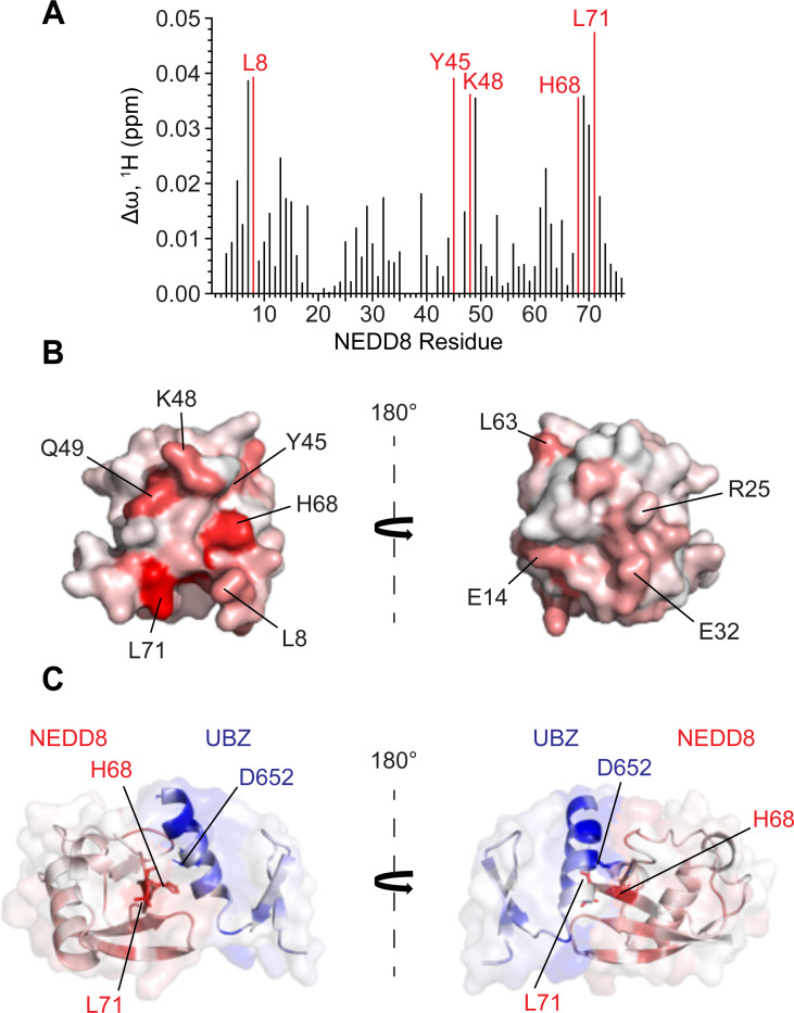
Figure 5: An NMR-based molecular model of the UBZ/NEDD8 complex.
(A) A bar graph of per-residue chemical shift perturbations (CSPs) in 15N-TROSY-HSQC spectrum of 15N-labelled NEDD8 caused by the addition of unlabelled UBZ domain of Pol η. Residues for which peaks disappeared due to broadening were assigned to the highest observed CSP value. (B) The UBZ domain binding site mapped on the surface of NEDD8 (PDB: 1NDD). The surface is color-coded according to the NMR CSPs from smallest (white) to largest (red). The structures are shown in two orientations with a 180° rotation (C) HADDOCK model of the UBZ domain of Pol η (blue) in complex with NEDD8 (red) colored according to their as in Figure 4B and 5C. The model is shown in two orientations with a 180° rotation.

