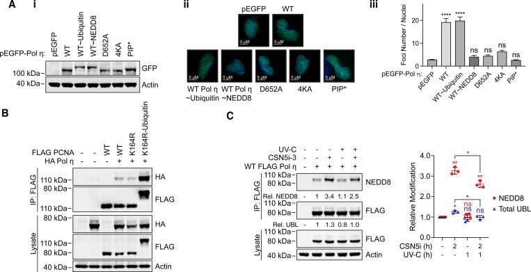
Figure 6: Mono-ubiquitination and mono-NEDDylation differentially regulate Pol η.
(A) The indicated GFP-Pol η variants were transiently expressed in MRC-5 cells. (i) The western blot demonstrates expression of each variant in whole cell lysates. (ii) Cells expressing each construct and growing on coverslips were exposed to 20 Jm2 of UV-C, fixed after 6 hours, and imaged on a fluorescence microscope with a 63x objective lens. Images taken in the EGFP channel were converted to 16-bit grayscale and the number of foci per nucleus measured. (iii) The graph represents the average number of foci per nucleus. Error bars represent standard deviation. Unpaired t-tests were used to assess differences between groups. **** = p <0.001, ns = not significant. (B) WT, K164R and a K164R-ubiquitin chimera of PCNA were immunoprecipitated from cells co-expressing HA Pol η. The lysate and eluent were immunoblotted as indicated. (C) 293T cells expressing WT FLAG Pol η were treated with 1 μM of the NEDDylation inhibitor CSN5i-3, or DMSO for 1 hour, before being exposed to 20 Jm2 of UV-C. Cells were harvested 1 hour after exposure, FLAG Pol η was immunoprecipitated, and the eluent and lysate immunoblotted as indicated. Relative mono-UBLylation was calculated based on the ratio of higher to lower FLAG Pol η bands. Mono-NEDDylation was calculated based on the ratio of NEDD8 to FLAG bands. The graph represents quantification from three independent repeats. Error bars represent standard deviation. Unpaired t-tests were used to assess differences in NEDDylation and UBLylation. * = p < 0.05, ** = p < 0.1, ns = not significant.

