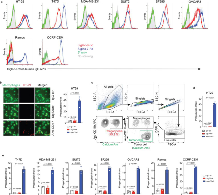
Extended Data Fig. 10 |. Siglec-7/9 degradation does not improve macrophage phagocytosis of cancer cells.
a, Flow cytometry analysis of the expression of Siglec-7 and Siglec-9 ligands on various types of tumor cell lines by staining with Siglec-7Fc and Siglec-9Fc, respectively. b, Fluorescence microscopy imaging-based measurement of hMDM (green) phagocytosis of pHrodo red-labeled HT29 cells in the absence or presence of Sig7/9de or anti-CD47 (blockade of the well-known “don’t eat me” signal CD47, which binds to signal regulatory protein-alpha (SIRPα) expressed on macrophages and dendritic cells (DCs) is used as the positive control). Scale bar, 50 μm. c, Gating strategy for flow cytometry-based phagocytosis assay, in which, phagocytosis was recorded by measuring the frequency of calcium-AM+ macrophages within the live CD11b+ population after removal of debris and doublets. d,e, Flow cytometry-based measurement of hMDM phagocytosis of the Siglec-7/9L+ cell lines of colon cancer (HT29), breast cancer (T47D and MDA-MB-231), PDAC (SUIT2), glioblastoma (SF295), ovarian cancer (OVCAR-3), B-lymphoma (Ramos) and T-ALL (CCRF-CEM) cancer cell lines, in the absence or presence of Sig7/9de or anti-CD47 (positive control). Data are mean ± s.d. Two-tailed unpaired Student’s t-test (b,d,e).

