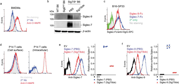
Extended Data Fig. 12 |. Siglec-7/9 degradation occurs in mouse immune cells.
a, The staining of M6PR of mouse BMDMs using anti-CI-M6PR. b, Western blot analysis of Siglec-7/-9 degradation in Sig7/9+ BMDMs in comparison to WT and SigEKO controls. c, Flow cytometry analysis of the expression of Siglec-7 and Siglec-9 ligands on B16-GP33 cells by staining with Siglec-7Fc and Siglec-9Fc, respectively. d, The staining of cell surface and intracellular M6PR on P14 T cells using anti-CI-M6PR. e,f, Analysis of cell-surface Siglec-7 (e)/-9 (f) depletion in Siglec-7 and Siglec-9 expressing P14 T cells upon treatment with 30 nM Sig7/9de for 1 hour.

