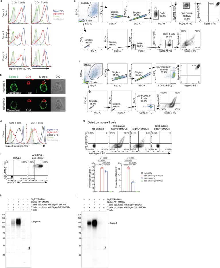
Extended Data Fig. 2 |. T cells acquire Siglec-7 and -9 molecules from interacting myeloid cells via trogocytosis.
a, Analysis of cell-surface expression of Siglec-7 and -9 ligands on CD8+ and CD4+ T cells from healthy donor PBMCs by staining with Siglec-7Fc and Siglec-9Fc, respectively. b, Representative fluorescence microscopy imaging analysis of Siglec-9 localization in interacting SEB-pulsed hMDMs and donor-matched PBMC T cells, which were pre-FACS sorted as Siglec-7/9– population. Scale bar, 10 μm. c, Gating strategy for flow cytometry-based quantification of Siglec-7/-9 transfer to T cells from hMDMs, in which, the frequency of Siglec+ T cells was measured within the CD3+ T cell population after removal of debris and doublets in the absence of hMDM cells. d, Flow cytometry analysis of cell-surface expression of Siglec-7, Siglec-9 and Siglec-E ligands on splenic CD8+ and CD4+ T cells from WT mice by staining with Siglec-7Fc, Siglec-9Fc and Siglec-E-Fc, respectively. e, Gating strategy for flow cytometry-based quantification of Siglec-7/-9 transfer to WT mouse T cells (CD45.1+/+) from Sig7/9+ mouse BMDMs (CD45.2+), in which, the frequency of Siglec+ T cells was measured within the CD45.1+ T cell population after removal of debris and doublets in the absence of BMDM cells. f, Flow cytometry analysis of the purity of CD3+ T cells after enrichment from CD45.1+/+ splenic T cells. g, Flow cytometry analysis of Siglec-7/9 trogocytosis to WT mouse T cells after 5-min coculture with Sig7/9+ BMDCs vs. SigEKO BMDCs with or without SEB pulsing. BMDC, bone marrow-derived dendritic cell. Data are mean ± s.d. Two-tailed unpaired Student’s t-test (g). h,i, Western blot analysis showing the transfer of intact Siglec-7 and -9 to WT mouse T cells from Sig7/9+ BMDMs compared to SigEKO BMDMs.

