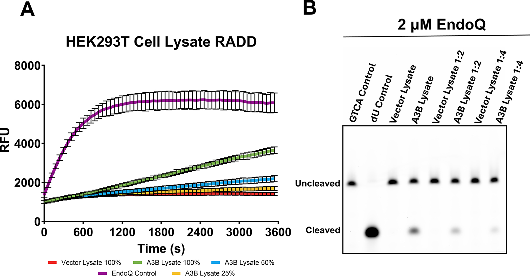
Fig. 7. Measuring A3B activity in HEK293T cell lysate.

(A) Real-time deamination readout of reactions containing various dilutions of A3B-expressing human cell lysate and controls transfected with empty vector (N = 3 for each condition). The trace in purple represents the EndoQ control reaction containing 1 μM TdU-ZEN, 2 μM EndoQ, and lysis buffer. The green, blue, and yellow traces represent 1 μM TC-ZEN reporter in the presence of undiluted, 1:2, or 1:4-diluted A3B-expressing cell lysate, respectively. The red trace represents the negative control, containing undiluted control cell lysate. All reactions contained 2 μM EndoQ. (B) After fluorescence scanning, the reactions were run on a 15% TBE-Urea gel. None of the control cell lanes showed any substrate processing (lower band) while the A3B-containing samples showed decreased substrate processing in-line with increased dilution.
Figure adapted from Belica, C., Carpenter, M. A., Chen, Y., Moeller, N., Harris, R. S., & Aihara, H. (2024). A real-time biochemical assay forquantitative analyses of APOBEC-catalyzed DNA deamination. Journal of Biological Chemistry.
