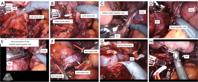
Figure 4.
The details of the surgery. (A) The left renal vein and medial IVC is dissected and exposed. (B) The right renal artery is secured caudal to the left renal vein, clipped, and taped. (C) The inferior right hepatic vein is dissected using Endo GIATM at of its confluence with the IVC. (D) The short hepatic vein is identified cephalad to the inferior right hepatic vein and is dissected using a LigasureTM. (E) Intraoperatively, an echo confirms that the tumor tip was slightly cephalic to the inferior right hepatic vein. (F) The IVC and left renal vein are clamped cephalad and caudally using laparoscopic bulldog forceps to enclose the tumor. (G) An incision is made on the IVC and the tumor thrombus is removed. Adhesion to the IVC wall is mild. (H) The IVC wall is sutured using 4-0 PROLENE sutures. IVC, inferior vena cava.

