
FIGURE 2.
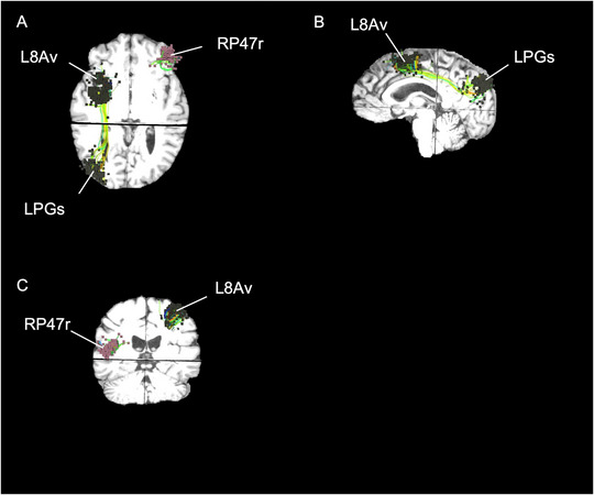
Anatomical locations of rTMS for Patient 1. (A) Axial, (B) sagittal, and (C) coronial visualization of rTMS targets superimposed on top of MRI of Patient 1. Patient 1 is a 48‐year‐old female with a diagnosis of MDD for 10 years. She had a baseline score of 8, posttreatment of 9, and follow‐up of 3 on the PSQI.