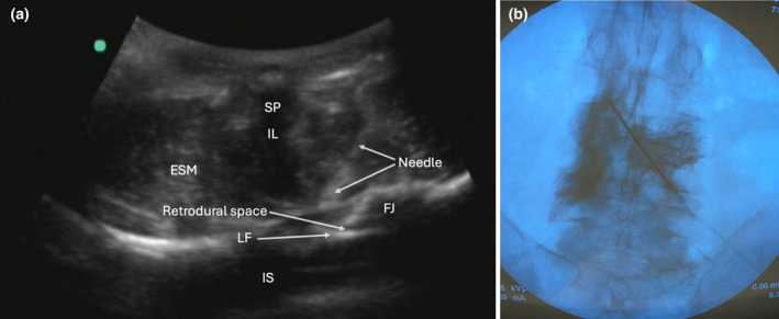
Figure 1.

(a) Ultrasound of the needle insertion and retrodural space. (b) Lumbar X‐ray anterior–posterior view with dye contrast spread in the retrodural space. ESM, erector spinae muscle; FJ, facet joint; IL, interspinous ligament; IS, intrathecal space; LF, ligamentum flavum; SP, spinus process.
