Figure 1.
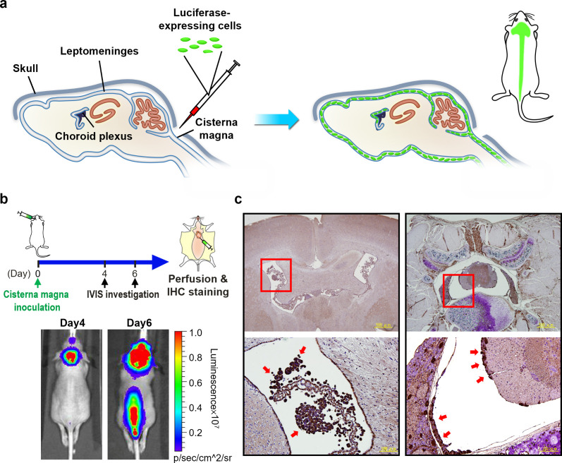
Establishment of a leptomeningeal carcinomatosis (LM) mouse model. (a) Scheme illustrating the direct inoculation of luciferase-expressing cells through cisterna magna to establish a LM orthotopic mouse model. (b) Monitoring of the mouse by IVIS. Prominent BLI signals over the brain and the spinal cord were observed at Day 6 post tumor cell inoculation, and the mouse was sacrificed for IHC confirmation. (c) Tumor cells (red arrows) observed in the ventricular space of the brain (left panel) and the spinal cord (right panel) in IHC studies.

