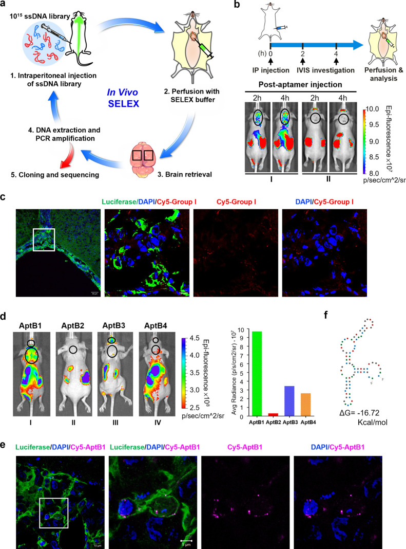
Figure 2.
Identification of AptB1, a BBB-penetrating and cancer-targeting aptamer by in vivo SELEX. (a) Scheme illustrating the process of in vivo SELEX. (b) The aptamers were divided into group I or II based on the QGRS prediction. The group I aptamer contained G-quadruplex structure and the group II did not. The LM mouse I administered with Cy5-labeled group I aptamers showed fluorescent signals over the brain/spine in a crescendo pattern at 2 and 4 h after injection. The lesions are outlined with black circles. (c) Confocal microscopy images revealing group I aptamer signals (red) within the tumor cells (green) on the leptomeninges. (d) Strong Cy5 fluorescent signals emitted from the brain and the spine were detected in the LM mouse administered with AptB1, as shown in the IVIS imaging and the quantified bar chart. The lesions are outlined with black circles. (e) The confocal microscopy images revealed AptB1 signals (pink) within the tumor cell (green) on the leptomeninges. (f) The Mfold prediction of AptB1 secondary structures.

