Figure 6.
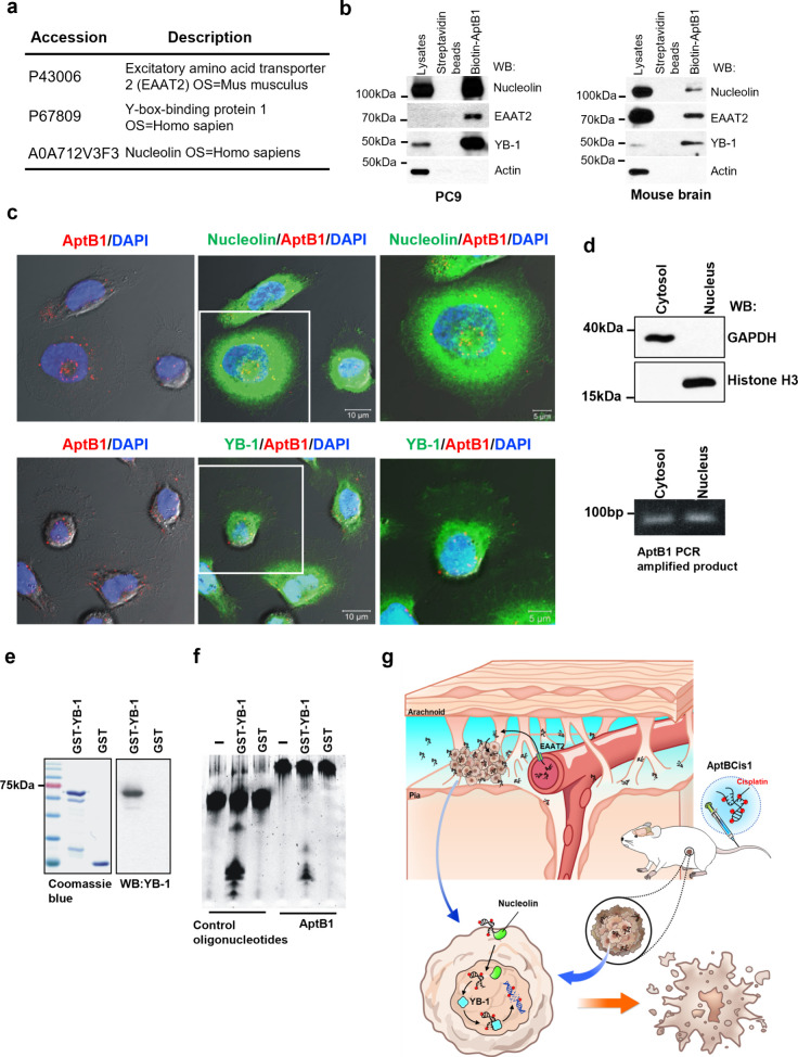
AptB1 interacting with EAAT2, Nucleolin, and YB-1. (a) Results of AptB1-AP/MS study revealed three candidate AptB1-interacting proteins: EAAT2, YB-1, and Nucleolin. (b) AP-immunoblots verified the interaction between AptB1 and EAAT2, Nucleolin, as well as YB-1 in the PC9 cells and in the mouse brain. (c) The confocal microscopy images showed colocalization (yellow) of AptB1 (red) and Nucleolin (green, upper panel) or YB-1 (green, lower panel) in the PC9 cells. (d) AptB1-treated cells were fractionated into cytosol and nucleus fractions. GAPDH is a cytosolic marker, and Histone H3 is a nucleus marker. The AptB1 sequences were successfully amplified in both cellular fractions. (e) The Coomassie Blue stain and the immunoblots showed the purified GST and GST-YB-1 proteins. (f) The YB-1 exonuclease assay results supported the role of YB-1 as an exonuclease for AptB1. (g) Scheme illustrating the proposed mechanism of AptBCis1 as therapeutics for lung cancer with and without LM.

