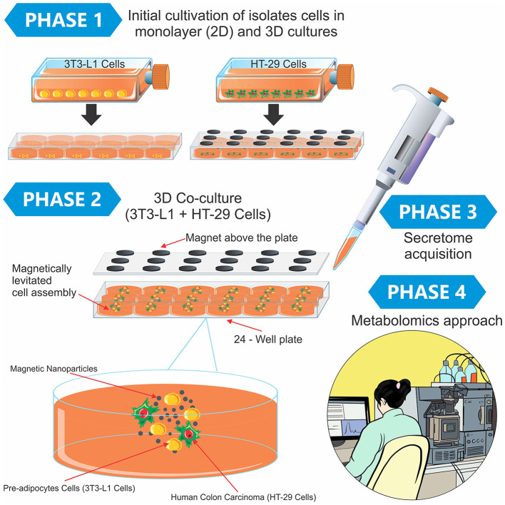
Figure 1.
Experimental design containing the phases of the study and a didactic illustration of the magnetic 3D cell culture. In Phase 1, the 3T3-L1 and HT-29 cells were separately cultured using the two-dimensional model in monolayer (2D) and 3D cultures. After this initial preparation, both cells (3T3-L1 + HT-29) were mixed in the 3D co-culture, in which the cells grew in spheroid geometry (Phase 2). Finally, the secretome was obtained from different cultures (Phase 3), and the metabolomics approach was conducted in Phase 4.

