FIG. 1.
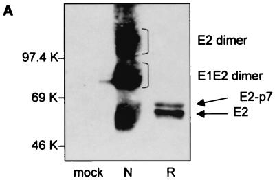
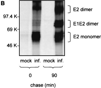
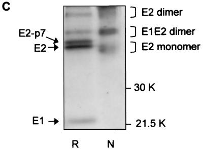
Analysis of BVDV envelope glycoproteins expressed in MBDK cells. (A) MBDK cells were infected with BVDV (NADL strain) at an MOI of 1. Eighteen hours p.i. the cells were lysed and analyzed by SDS–10% PAGE under nonreducing (lane N) and reducing (lane R) conditions, followed by Western blot analysis with MAb 214. (B) MBDK cells were infected with BVDV at an MOI of 1. Eighteen hours p.i. the cells were pulse-labeled with [35S]methionine and [35S]cysteine for 15 min, chased for the times indicated, lysed, and immunoprecipitated with MAb 214. Immunoprecipitated proteins were separated by SDS–10% PAGE under nonreducing conditions and analyzed by autoradiography. (C) Conditions were the same as described for panel B, except that the sample at min 90 of chase was analyzed by SDS–12% PAGE, under both nonreducing and reducing conditions. Mock-infected cells (mock inf.) were included as controls.


