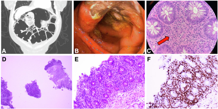
Fig. 1. Representative images of IEC-associated enterocolitis.
Top panel: Representative colitis-related images from different patients. Panel A: diffuse bowel wall pneumatosis and pneumoperitoneum on computed tomography imaging consistent with toxic megacolon, which subsequently required subtotal colectomy. Panel B: ulcerations noted on endoscopic evaluation of the transverse colon. Panel C: 10× magnification of colonic mucosa showing focal crypt dropout and increased apoptotic activity; minimal expansion by scattered eosinophils and focal intraepithelial lymphocytes are also seen. Bottom panel: Representative enteritis-related images from one patient. Marked infiltration of the lamina propria by T lymphocytes at low power (2×, panel D) and high power (10×, panel E). Immunohistochemical stain for CD3 (20×, panel F) demonstrates that the majority of the lamina propria cells and intraepithelial lymphocytes are T cells.

