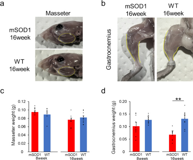
Fig. 3.
Muscle Weight Analysis (a) The masseter muscle in mSOD1 and WT groups at 16 weeks of age, respectively. (b) The gastrocnemius muscle in mSOD1 and WT groups at 16 weeks of age, respectively. (c) The wet weight of the masseter muscle in mSOD1 and WT groups at 8 and 16 weeks. There were no significant differences observed between the groups. (d) The wet weight of the gastrocnemius muscle in mSOD1 and WT groups at 8 and 16 weeks. At 16 weeks, the mSOD1 group had significantly lower values compared to the WT group [8 weeks (mSOD1: n = 5, WT: n = 5), 16 weeks (mSOD1: n = 5, WT: n = 5), as assessed by Student’s t-test, p < 0.01, **p < 0.01].

