. 2024 Oct 3;15(44):18443–18454. doi: 10.1039/d4sc00402g

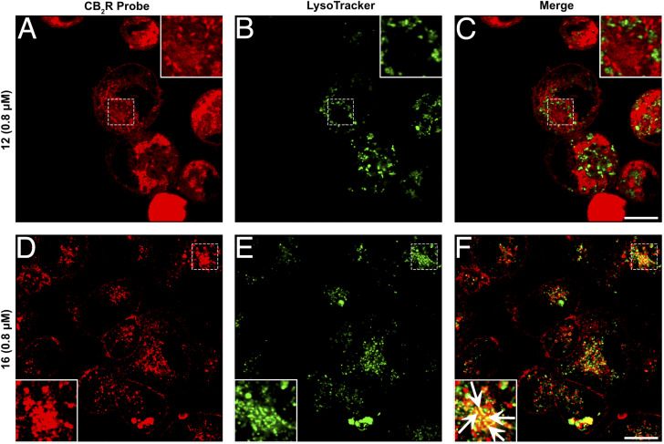
Fig. 6. Co-staining studies utilizing super-resolution confocal microscopy images of hCB2R-CHO cells. (A–F) Double-staining of hCB2R-overexpressing CHO cells exposed for 15 min with 12 (A) or 16 (D) together with LysoTracker, a lysosome marker (B and E, green). Merged images are shown in the panels (C) and (F). The superimposition of the two stainings revealed that 12 and LysoTracker did not colocalize, while 16 and LysoTracker partially co-stained in several dot structures (white arrows in the magnified box). The images are representative of three independent experiments, and in each case, five fields were examined. Scale bars, 10 μm.

Fig. 6