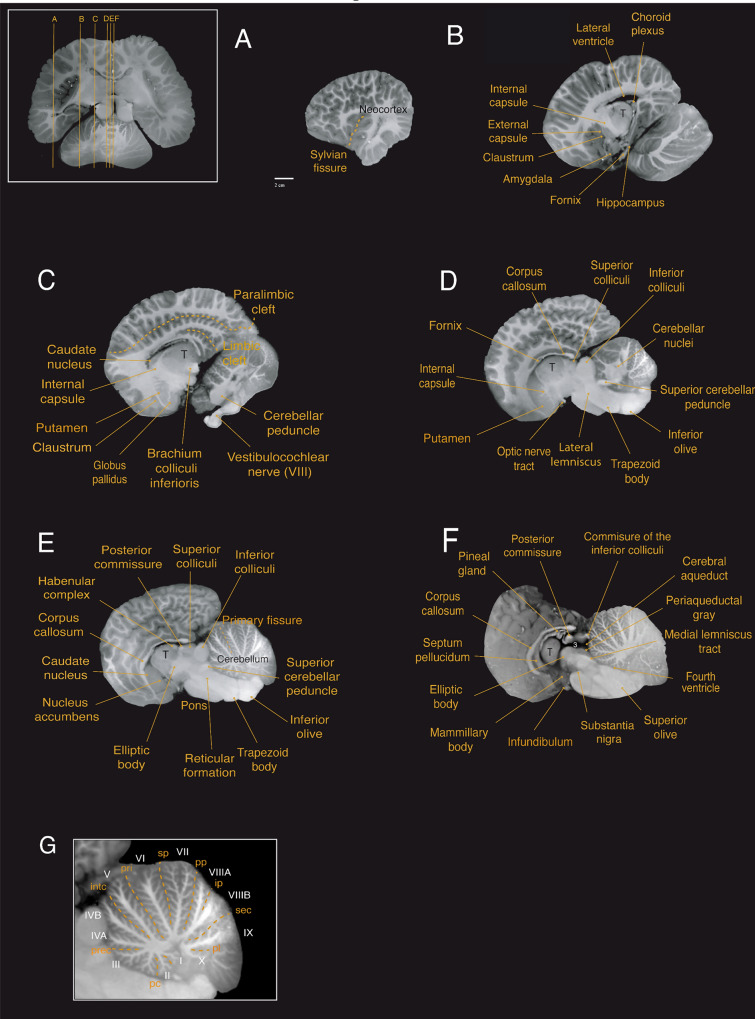
Fig. 5.
Sagittal magnetic resonance imaging (MRI) scans of the Guiana dolphin brain at 1.9 mm interval—from to axis; T2 weighted; MRI grayscale inverted. Scale bar = 2 cm. Top left: Illustration of the position of the brain in the horizontal plane. In the bottom, the midsagittal cerebellar section demonstrates the lobules and arborization patterns of the vermis of the structure. Cerebellar lobules are labeled in white, and sulci are labeled in yellow. For abbreviations see list

