
FIGURE 4.
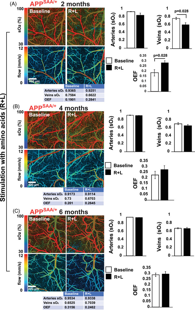
Amino acid‐induced oxygen consumption is blocked in the APPSAA/+ mouse brain. A–C, MP‐PAM imaging of APPSAA/+ mouse cortex through an open‐skull window before and 80 minutes after a topical application of amino acids (R+L). Decreases in cortical vasculature sO2 and OEF were observed only in the 2‐month‐old group. sO2 in arteries, veins, and OEF values for the experiments shown in the figure are depicted in the table below each image. The bar graphs show the quantification of four independent experiments. Statistical analyses were performed using Student two‐tailed unpaired t test. Error bars represent ± standard error of the mean. Four animals/age groups were used in these experiments. APPSAA/+, heterozygous amyloid precursor protein knock‐in; MP‐PAM, multi‐parametric photoacoustic microscopy; OEF, oxygen extraction fraction; R+L, arginine and leucine; sO2, saturation of hemoglobin.