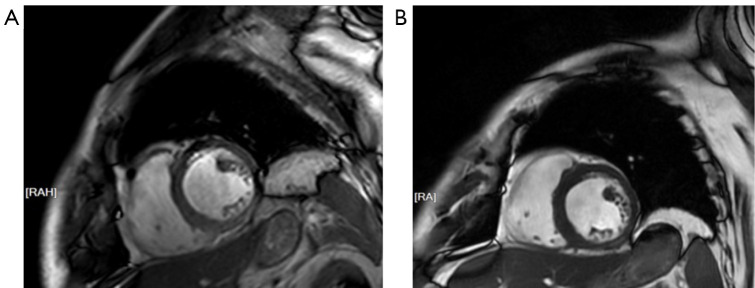
Figure 1.
CMR images of the patient before and after treatment in our department. (A) The thickness of midventricular inferior and inferolateral wall seemed to be slightly lower than normal. (B) The thickness of corresponding ventricular appeared normal in subsequent follow-up. RA, right anterior; RAH, right anterior head; CMR, cardiac magnetic resonance.

