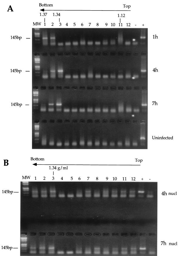
FIG. 1.
PCR analyses of cytoplasmic (A) and nuclear (B) extracts after equilibrium density fractionation using primers specific for the strong-stop DNA (expected band size is 145 bp). HeLa cells were infected with an HIV-1-based vector; cell extracts were prepared 1, 4, and 7 h postinfection, loaded on a 20 to 70% linear sucrose gradient, and centrifuged at 4°C for 20 h at 35,000 rpm in a Beckman SW55 rotor. After centrifugation, gradients were collected in 12 fractions and analyzed. Arrows indicate the direction of the gradient from the lowest (top) to the highest (bottom) density. The density of the fraction containing the peak of the viral DNA is indicated for each time point. Lanes: MW, DNA molecular weight standards; 1 to 12, fractions 1 to 12. The rapidly migrating bands are PCR artifacts. Hirt viral DNA was used as positive control (+); uninfected Hela cells served as a negative control (−). nucl, nuclear.

