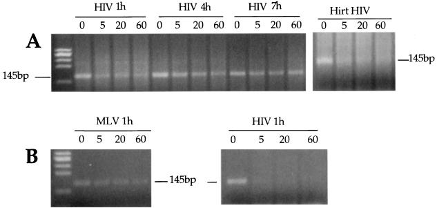
FIG. 7.
Sensitivity of the viral strong-stop DNA within the RTC to micrococcal nuclease digestion. (A) Equilibrium density fractions containing the peak of the viral DNA from the cytoplasmic extracts collected 1, 4, and 7 h postinfection were incubated on ice in the presence of micrococcal nuclease and 2 mM CaCl2. The reactions were stopped by addition of 4 mM EGTA at the indicated time points and analyzed by PCR using primers specific for the strong-stop DNA (expected band size is 145 bp). Naked viral DNA was incubated as above in the presence of micrococcal nuclease and cytoplasmic extracts from uninfected cells (Hirt HIV). (B) Equilibrium density fractions containing the peak of HIV-1 and MoMLV DNAs were mixed 1:1 and subjected to micrococcal nuclease digestion as above. The reaction was analyzed by PCR using primers specific for HIV or MoMLV strong stop. To ensure the linearity of amplification, the samples were subjected to two independent amplification rounds, one of 30 and one of 40 cycles.

