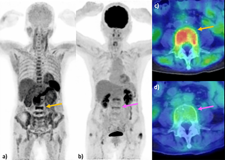
Figure 1. Comparison of PET imaging in MM: 11C-acetate versus 18F-FDG.
Imaging results from a 68-year-old woman with MM are presented. MIP images reveal multiple lesions in the vertebrae, including the L5 vertebral body (arrow) and pelvic bones, identified using 11C-acetate PET (a). Lesions in the lumbar vertebrae, including the L5 vertebral body (arrow), are detected by 18F-FDG PET (b). An axial fused PET/CT image utilizing 11C-acetate at the L5 vertebra demonstrates increased uptake in the vertebral body (SUVmax 4.0) (arrow) (c), in contrast to the relatively weak uptake observed in the 18F-FDG PET/CT image (SUVmax 2.0) (arrow) (d).
FDG, fluorodeoxyglucose; MIP, maximum intensity projection; MM, multiple myeloma; SUV, standardized uptake value

