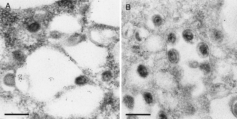
FIG. 4.
Immunolabeling of TGN38 protein. Ultrathin sections of Lowicryl-embedded PrV-infected porcine kidney cells were processed as described in Materials and Methods and incubated with two different dilutions (A, 1:100; B, 1:1,000) of anti-TGN38 serum (44) followed by 10-nm-diameter gold particle-tagged secondary antibodies. The bars represent 250 nm.

