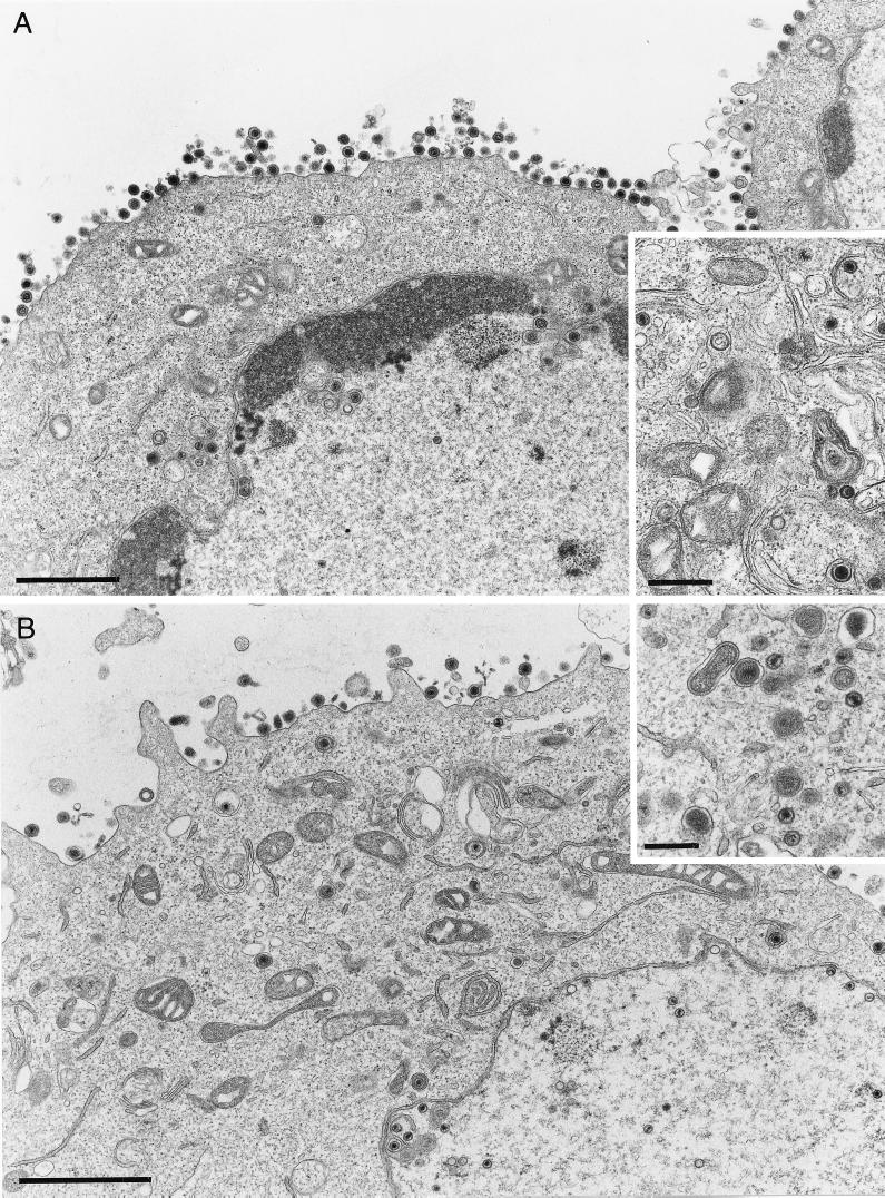
FIG. 6.
Ultrastructure of cells infected with gB or gH deletion mutants of PrV. Noncomplementing RK13 cells were infected with PrV-gB− (A) or PrV-gH− (B) at an MOI of 1 and analyzed 16 h after infection. All stages of virus maturation, including numerous extracellular virus particles, can be observed. The bars represent 1.5 μm in panels A and B and 500 nm in the insets.

