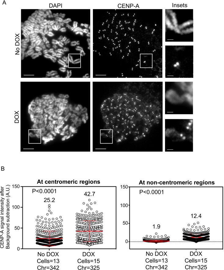
Figure 2.
β-TrCP depletion contributes to CENP-A mislocalization in MDA-MB-231 cells. (A) Representative images of metaphase chromosome spreads prepared from MDA-MB-231Δ β-TrCP cells untreated or treated with DOX for 48 h, showing the localization of endogenous CENP-A on mitotic chromosomes. Scale bars: 5 µm for main images, 2 µm for insets. (B) Quantification of CENP-A signal intensities (arbitrary units) at centromeric (left) and noncentromeric (right) regions in metaphase chromosome spreads of MDA-MB-231Δ β-TrCP cells treated as in B. Each circle represents one spot quantified on chromosome. “Chr” represents number of chromosomes analyzed in the number of cells denoted. Error bars depict the SD across areas measured in the number of cells as indicated from three biological repeats and the P-values were calculated using Student’s t test.

