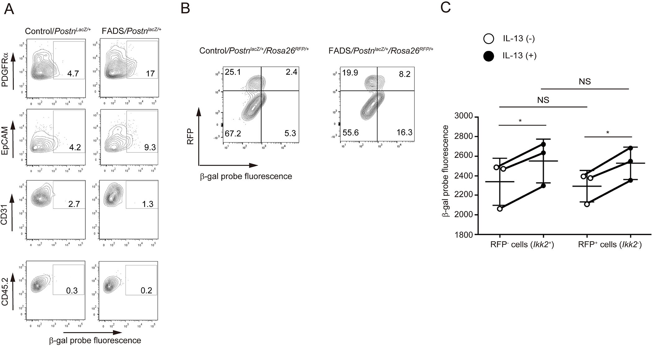
Figure 2. Identification of periostin-producing cells in the skin lesions of FADS mice.

(A) β-galactosidase (β-gal) positive cells in PDGFRα+ and EpCAM+ facial dermal cells of 4-week-old FADS/PostnlacZ/+ mice and control/PostnlacZ/+ mice. PDGFRα+-, EpCAM+-, CD31+-, and CD45.2+-gated cells are depicted. (B) β-gal positive cells in both RFP+PDGFRα+ and RFP−PDGFRα+ cells derived from 4-week-old FADS/PostnlacZ/+/Rosa26RFP/+mice and control/PostnlacZ/+/Rosa26RFP/+mice. (C) β-gal expression by treatment with or without 50 ng/mL IL-13 for 24 hours in both RFP− (Ikk2+) and RFP+ (Ikk2−) primary cultured dermal fibroblasts. The data shown are the mean ± SEM of three fibroblast samples prepared separately from three mice. Similar results were obtained from three separate experiments. Statistical analysis was performed using a two-sided, paired Student t-test; *P < 0.05, **P < 0.01. NS, not significant.
