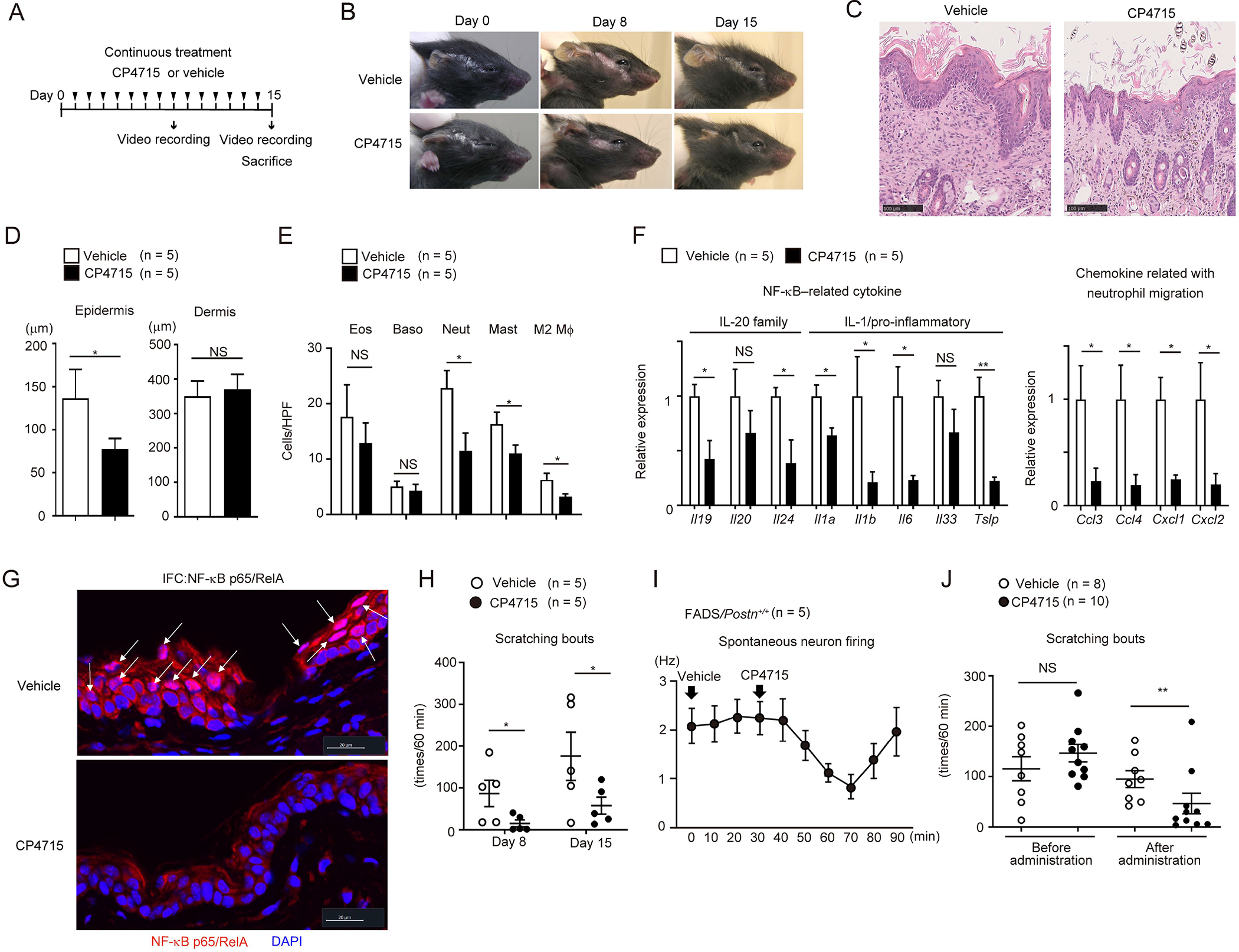
Figure 6. Effects of pharmacologically inhibiting periostin on skin inflammation and itching in FADS mice.

(A) The experimental protocol of continuous CP4715 administration. (B-H) Macroscopic facial skin lesions (B), HE staining (C), epidermal and dermal thickness (D), the numbers of per high-power field (HPF: ×40) of the indicated inflammatory cells, expression of the indicated cytokines and chemokines (F), p65 immunostaining (G), and scratching bouts (H) of 3-week-old FADS/Postn+/+ mice treated with continuous administration of vehicle (n = 5) or CP4715 (60 mg/kg) (n = 5) on day 0, day 8, and day 15 (B), or on day 15 (C-G), or on day 8 and day 15 (H). Scale bars: 20 (G) or 100 μm (A). White arrows indicate epidermal cells with nuclear p65 expression (G). (I, J) Spontaneous firing of dorsal horn neurons (I) and scratching bouts (J) in 60 minutes after 10–14-week-old (I) (n = 5) or 4-week-old (vehicle, n = 8; CP4715, n = 10) (J) FADS/Postn+/+ mice were treated with single-shot administration of vehicle or CP4715 (30 mg/kg, see also Figures S5 and S6). The data shown are the mean ± SEM of samples obtained from three (D–G), five (I), or four (J) independent experiments. Statistical analysis was performed using a one-sided Mann-Whitney U-test; *P < 0.05, **P < 0.01. NS, not significant.
