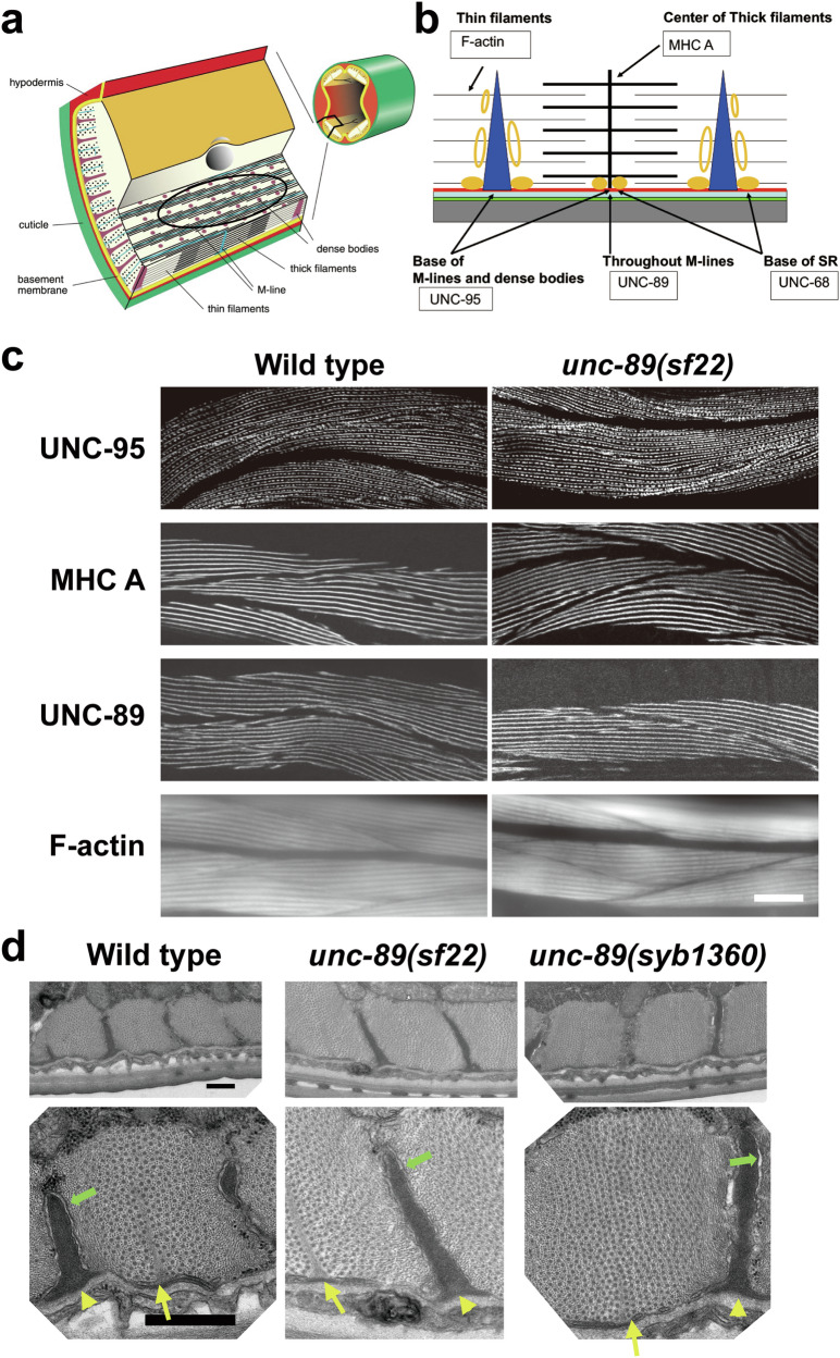
Fig. 2. Nematodes expressing UNC-89 with the KtoA mutation in PK2 have normal muscle structure.
a The drawing on the right shows a cross-section through an adult worm indicating that the body wall muscle is organized into four quadrants. Each quadrant is composed of interlocking pairs of mononuclear fusiform-shaped cells (23 or 24 per quadrant). The enlargement emphasizes that the myofilament lattice is restricted to one side of the cell. The drawing depicts several planes of section; one plane emphasizes that this is striated muscle with typical A-bands containing thick filaments organized around M-lines, and overlapping thin filaments attached to dense bodies (Z-disk analogs). The black oval indicates the plane viewed when an animal, lying on a glass slide, is examined by light microscopy as shown in part (c). This drawing is a slight modification of a published drawing2, under terms of a Creative Commons Attribution License. b Idealized sarcomere and SR with the location of proteins localized in part (c). MHC A: myosin heavy chain A. Red: muscle cell membrane; blue-green: ECM; green: hypodermis; and grey: cuticle. Orange ovals: location of SR determined by electron microscopy; solid orange ovals: location of UNC-68 (Ryanodine receptor). c Wild type (left column), and unc-89(sf22) (right column) animals immunostained with various antibodies to proteins indicated in part (b). Phalloidin was used to image F-actin in thin filaments. In each panel, several spindle-shaped body wall muscle cells are shown. Note that there is no difference in the pattern of localization of these sarcomeric markers between wild type and unc-89(sf22). Scale bars, 10 μm. d Transmission EM of body wall muscle from wild type, unc-89(sf22) and a second independently-generated PK2 KtoA mutant, unc-89(syb1360). Bottom row displays the enlargement of one portion of the image above it. Yellow arrows, M-lines; yellow arrowheads, dense bodies, green arrows, SR. There is no discernible difference between the wild type and the two mutants in terms of numbers and organization of thick and thin filaments, M-lines, dense bodies, and SR. Scale bars, 0.5 μm.

