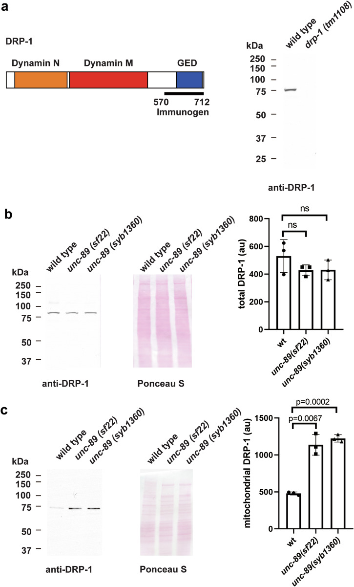
Fig. 6. More DRP-1 is associated with UNC-89 PK2 KtoA mutant mitochondria as compared to wild-type mitochondria.
a Left: Schematic representation of domains in DRP-1 as predicted by PFAM, and location of the immunogen used to raise antibodies to DRP-1. Right: Western blot showing the reaction of affinity-purified anti-DRP-1 to a protein of expected size for DRP-1 from wild type, and absence of detectable DRP-1 protein from the drp-1 null mutant. b Left: A representative western blot showing the reaction of anti-DRP-1 against total protein extracts from wild type, unc-89(sf22), and unc-89(syb1360). Right: quantitation of results indicating no significant difference in total DRP-1 levels in wild type vs. the two unc-89 mutants. au: arbitrary units; ns: not significant. Statistical significance was assessed using an unpaired t-test with Welch’s correction. c Left: A representative western blot showing the reaction of anti-DRP-1 against extracts of mitochondrial preparations from wild type, unc-89(sf22), and unc-89(syb1360). Right: quantitation of results indicating that there is more DRP-1 associated with mitochondria in the two mutants vs. wild type. au: arbitrary units. Statistical significance was assessed using an unpaired t-test with Welch’s correction.

