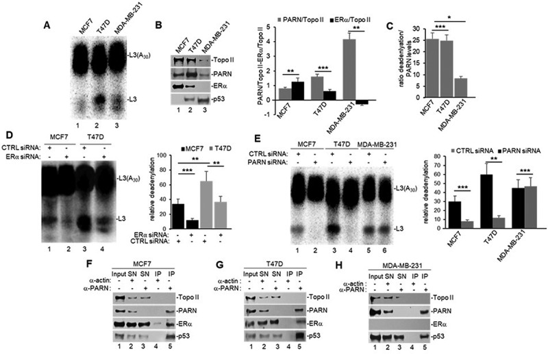
Figure 2.

ERα activates PARN-mediated nuclear deadenylation in (ERα+) cells (MCF7 and T47D), but not (ERα-) cells (MDA-MB-231). A) in vitro deadenylation assay and B) Western blot analysis using NEs from untreated breast cancer cells, prepared and analysed as in Figure 1A. Quantifications of PARN or ERα expression in B) is shown in a bar graph and normalized to Topo II loading control. C) normalization of PARN-mediated deadenylations shown in A) to PARN protein levels shown in B) for each cell line. D) in vitro deadenylation assay using NEs from cells treated with either CTRL or ERα siRNA for 24 h were performed as in 2A. E) in vitro deadenylation assay using NEs from breast cancer cells treated with either CTRL or PARN siRNA were analysed as in 2A. F–H) NEs from breast cancer cells were used in endogenous co-IP assays with anti-PARN and IgG antibodies, and performed as in Figure 1E. All figures show representative deadenylation reactions and Western blot analyses from at least three independent biological assays analysed by triplicate (n = 3). Experiments with two groups were analysed using two-tailed unpaired Student’s t-test. The p-values are indicated as *(<0.01) and ***(<0.0001).
