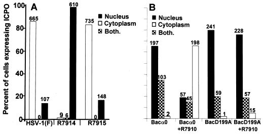
FIG. 1.
The requirements for the translocation of ICP0 from the nucleus to the cytoplasm. (A) ICP0 carrying the D199A substitution encoded by R7914 is not translocated to the cytoplasm late in infection of HEL fibroblasts. Replicate slide cultures of HEL fibroblasts were infected with 20 PFU of HSV-1(F), the recombinant virus R7914, or the repaired virus R7915 per cell and maintained at 37°C. At 12 h after infection, the cells were fixed and reacted first with polyclonal rabbit serum directed against exon II of ICP0 and second with FITC-conjugated goat anti-rabbit immunoglobulin antibodies. Sequential fields were examined in a Zeiss confocal microscope, and the numbers of cells exhibiting nuclear, cytoplasmic, or both nuclear and cytoplasmic localization of ICP0 were tabulated as shown in the histogram. The numbers above the bars indicate the numbers of cells showing a specific distribution of ICP0. (B) A viral gene function mediates the translocation of ICP0 from the nucleus to the cytoplasm during productive infection. Replicate slide cultures of HEL fibroblasts were exposed first to recombinant baculoviruses encoding wild-type or mutant ICP0 (D199A). After 2 h, cells were treated with 5 mM Na-butyrate. At 3 h after exposure to baculoviruses, the cells were exposed to the recombinant virus R7910, from which both copies of the α0 gene had been deleted. After 15 h of exposure to baculoviruses alone or 12 h after infection with R7910, the cells were fixed and reacted with rabbit polyclonal antibody against ICP0 and mouse monoclonal antibody against gD and then reacted with FITC-conjugated goat anti-rabbit IgG and Texas Red-conjugated goat anti-mouse IgG antibodies. Cell counts were done as described above, except that only cells exhibiting gD encoded by R7910 and ICP0 encoded by baculoviruses were counted.

