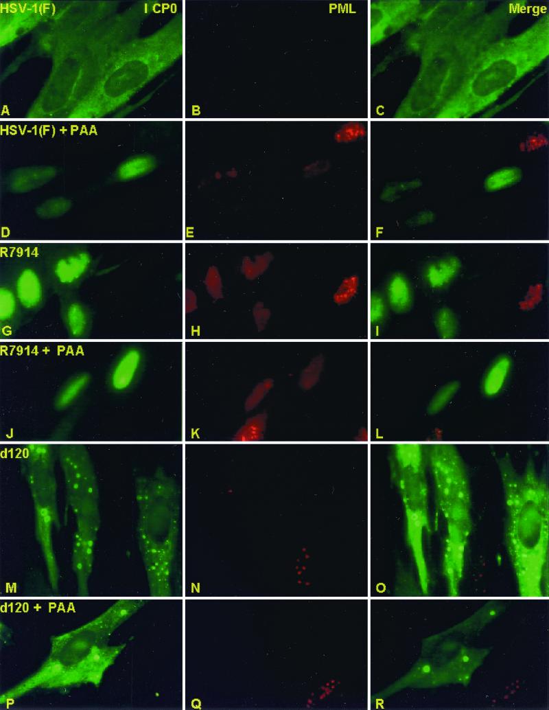
FIG. 3.
Immunofluorescent images of HEL fibroblasts infected with HSV-1(F), R7914, or d120 mutant and either treated or not treated with PAA. The images shown are representative of infected cells treated, generated, and counted as described in the legend to Fig. 2. The cells were reacted with rabbit polyclonal antibody to ICP0 and mouse monoclonal antibody to PML and then reacted with goat anti-rabbit IgG conjugated to FITC and goat anti-mouse IgG conjugated to Texas Red. The left, middle, and right columns show the cell localization of ICP0 and PML and merged images, respectively. The images were captured with a Zeiss confocal microscope with the aid of software provided by the manufacturer.

